|
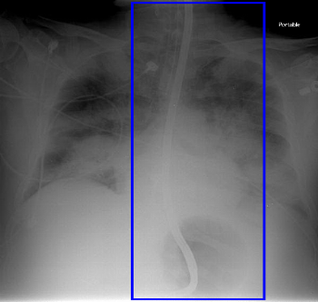
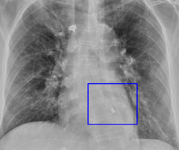
Feeding Tube
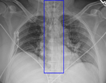
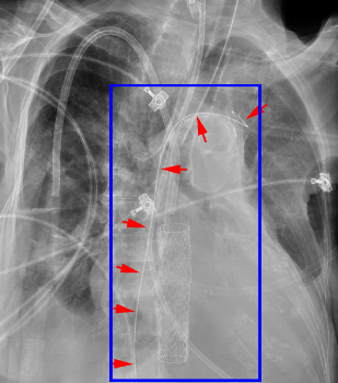
The feeding tube, often referred to as Dobhoff after its inventors, is slightly different from the nasogastric tube in that it is typically used for feeding (hence the name). It has a small opening at the side of the tip, which is radiopaque. You may also see the stiffening wire, as in this example, although that is removed before use.
|
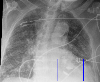
Mediastinal Drain
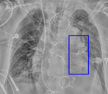
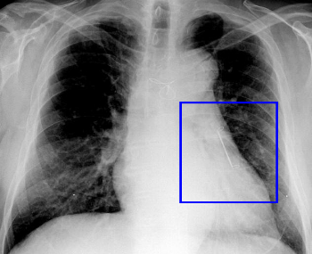
Postoperative drain, usually of the same type as the chest tube.
|
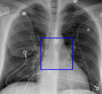
Minnesota Tube
Balloon-tipped nasogastric tube used to tamponade gastric variceal bleeding. The Blakemore tube is a closely related device with slightly different design.
|
|
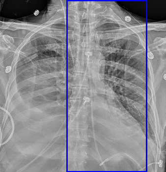
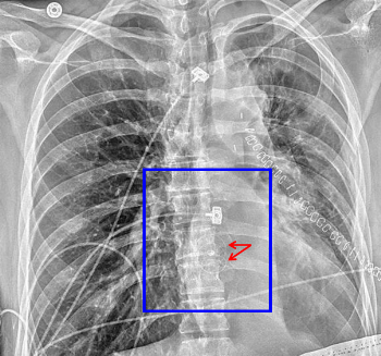
Nasogastric Tube
Tube generally used to drain/decompress the stomach. It has both an end-hole and a side-hole, both of which should be placed within the stomach.
|
|
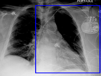
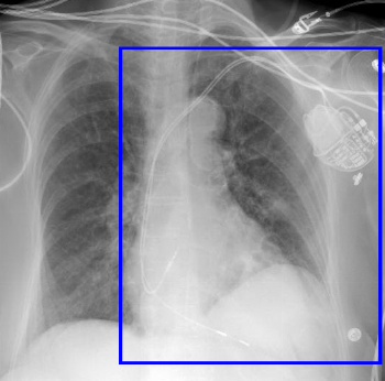
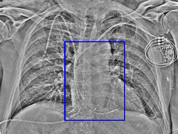
Biventricular Pacemaker
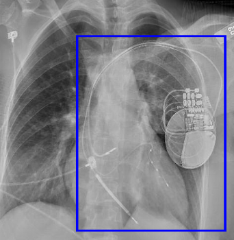
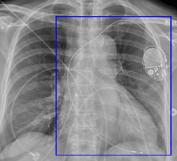
Leads in the right ventricle and a coronary vein (pacing the left ventricle). Typically these also have a right atrial lead, but they do not have to, such as in the example shown.
|
Dual Chamber Pacemaker
Two leads: right atrium and right ventricle.
|
Dual Chamber Pacemaker with Bundle of His Lead
The bundle of His lead is intended to provide more physiologic pacing of the ventricles. It is placed in the superior atrioventricular septum (level of the AV valves in the right heart).
This patient has conventional RA and RV leads as well.
|
|
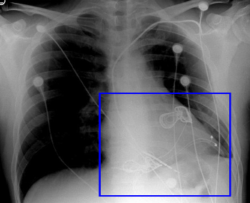
Epicardial Defibrillator Patch
If transvenous access has failed, defibrillator leads can be placed epicardially as well. These are referred to as patches given their broad, net like shape.
In this patient, you can see 2 patches as well as epicardial sensing leads. The pulse generator is below the field of view. (An abandoned transvenous defibrillator lead is seen as well.)
|
Epicardial Pacemaker
Epicardial leads are typically placed if transvenous access has failed. Temporary epicardial pacing leads are frequently used after cardiac surgery; however they are very much thinner and fainter on imaging.
|
Esophageal Temperature Probe
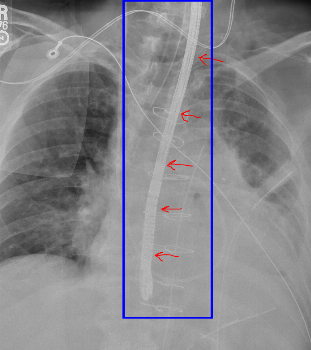
Braided-wire probe with radiopaque tip providing internal temperature monitoring.
|
|
Extravascular Defibrillator
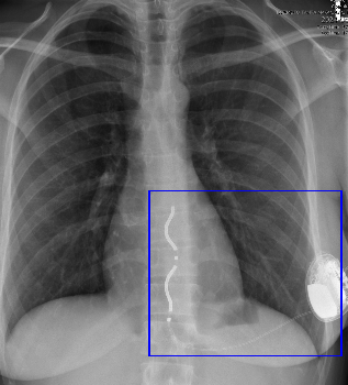
The extravascular defibrillator is in many ways similar to the subcutaneous defibrillator, but the lead is loculated posterior to the sternum in the anterior mediastinum. The lead also generally has a serpentine shape.
|
Impella
Impella LVAD is placed via a femoral or axillary arterial approach. It is a temporary ventricular assist device.
|
Implanted Defibrillator
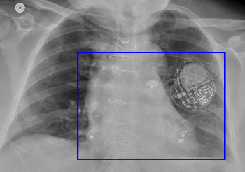
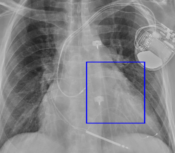
Implanted Cardioverter-Defibrillator (ICD) contains pacemaker and defibrillator functionality. It may have single chamber, dual chamber, or (often) biventricular leads. The leads in an ICD are characterized by one or more coils - a thickened portion that is used to deliver the shock. The pacemaker 'can' (pulse generator) is also distinguished by the presence of a capacitor as well as a battery.
The defibrillator component requires 2 coils; sometimes both coils are along a lead or leads in the venous system, but now the can itself can serve as one lead.
|
|
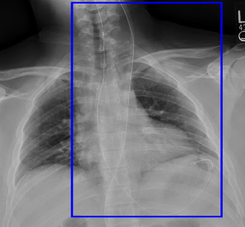
Intra-Aortic Balloon Pump
(aka IABP) Used for circulatory support, the balloon inflates in diastole and deflates in systole. Generally, only the radiopaque tip of the catheter is visualized, although if it is caught while inflated, you may see the balloon itself (over the descending aorta). The balloon is quite long and extends for the majority of the descending aorta's length.
The ideal position of the balloon is just distal to the subclavian artery and just proximal to the celiac artery, so that it does not obstruct either. General rules of thumb are either just below the aortic knob or 2 cm above the carina (see PMID 17717232).
Note that some IABPs are now being placed via an axillary approach. For these, the marker is usually left at the subclavian artery origin on purpose.
|
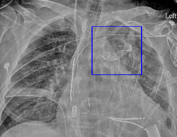
Retained Guidewire
The guidewire used to place a central line using Seldinger technique may be inadvertently left behind after central line placement. These wires often have a 'J' tip, as in this case.
Note that this wire enters from the IVC, likely a femoral central line, and terminates in the left brachiocephalic vein.
|
Single Chamber Pacemaker
Single lead typically in the right ventricle.
|
|
Swan-Ganz Catheter
Also known as pulmonary artery catheter. Used to monitor pulmonary artery and pulmonary capillary wedge pressures.
The tip of the catheter should be no farther than the hilum; typically it is left in the RVOT, main PA, or branch PAs. If it migrates too distally, there is a chance it could perforate the small pulmonary artery branch.
|
Transesophageal Echo Probe (TEE)
Used only intraoperatively to monitor cardiac surgery.
|
|
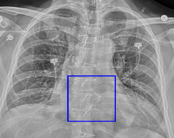
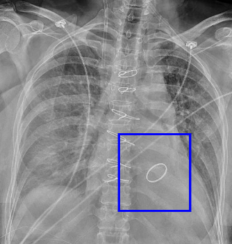
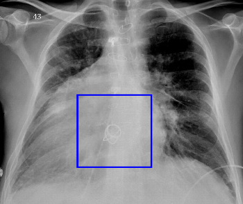
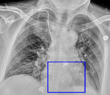
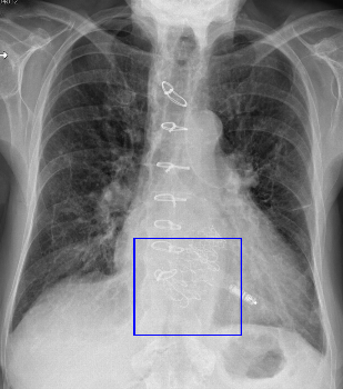
CABG Ring Marker
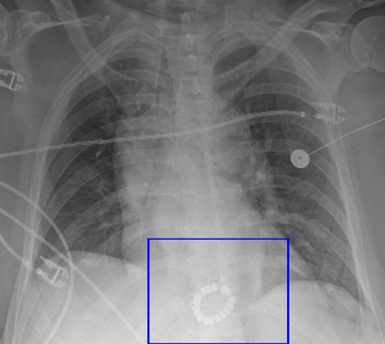
Rings mark the origins of coronary artery bypass grafts from the aorta. These provide a guide for angiographers.
This patient has two.
|
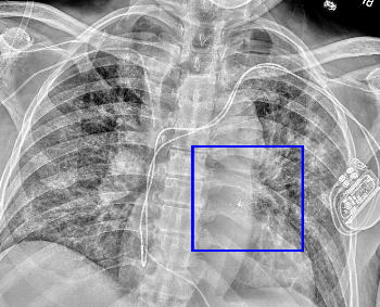
CardioMEMS
Wireless pressure sensor placed in a pulmonary artery branch (typically left lower lobe). Used for remote monitoring of heart failure patients.
|
Dual Leadless Pacemaker
This is a dual chamber leadless pacemaker, brand name Aveir. It is composed of two devices, one in the right atrium and the other in the right ventricle.
|
|
Leadless Pacemaker
Currently sold under the brand name Micra (others will likely follow). This can appear very similar to the chest wall loop recorder; the lateral radiograph will be helpful to confirm intracardiac placement (within the RV) versus chest wall placement.
|
MitraClip
Endovascular treatment for mitral regurgitation. This clip holds the leaflets together.
|
TEER
TEER (Transcatheter Edge-to-Edge Repair) is another brand of mitral valve clip device.
|
|
Wireless Esophageal pH Probe
|
|
Amplatzer ASD Closure Device
Endovascularly placed device to occlude an atrial septal defect. Appears as 2 faint circular objects connected by a small radiopaque line (these lie on either side of the defect). May be used to occlude VSDs and other holes, as well.
|
Annuloplasty
Annuloplasty rings are used to narrow and reform the shape of a dilated, regurgitant valve. These are typically used for the mitral and tricuspid valves. They are often distinguished by an incomplete radiopaque ring.
This is a tricuspid annuloplasty.
|
AtriClip
Left atrial appendage clip to occlude the appendage in patients with atrial fibrillation (to prevent embolism of thrombus).
|
|
Linx
Anti-reflux ring for the distal esophagus
|
Percutaneous Tricuspid Valve
This is an Evoque percutaneously placed tricuspid valve prosthesis
|
Prosthetic Aortic Valve
May have mechanical or biologic leaflets. This valve is bioprosthetic.
|
|
Prosthetic Mitral Valve
May have biologic (bioprosthetic) or mechanical leaflets. This example is a mechanical valve, although the leaflets are not clearly visible.
|
Starr-Edwards Mechanical Valve
Older variety of mechanical valve with ball and cage design. This example is an aortic valve prosthesis in a patient with DORV and dextrocardia.
|
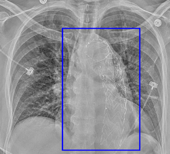
Transcatheter Aortic Valve Replacement
also known as TAVR or TAVI. Endovascularly placed bioprosthetic aortic valve for aortic stenosis. There are several different models but all have an outer wire mesh (stent) and radiolucent leaflets.
|
|
Transcatheter Tricuspid Valve
This is the Evoque transcatheter tricuspid valve prosthesis. The patient also has a TAVR.
|
Watchman
Left atrial appendage occluder (the cage plugs the opening of the appendage).
|
|
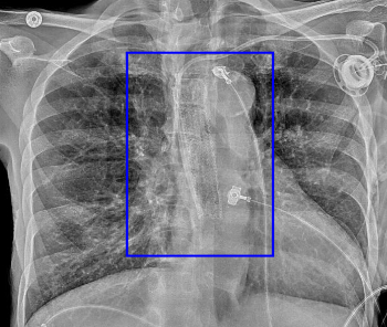
Intravascular Ventricular Assist Device
Currently the brand name is NuPulse iVAS. This is essentially a long-term implanted intra-aortic balloon pump. It includes several subcutaneous ECG leads in the chest wall; an axillary radiopaque implanted sheath; and a single radiopaque marker for the top of the balloon.
|
Laparotomy Sponge
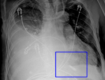
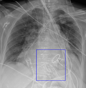
Surgical sponges have a radiopaque band to help identify them. They may be placed for packing an open wound or retained inadvertently after surgery. The sponge marker is often broad, whereas gauze has small crinkled lines.
This patient has an open sternotomy and biventricular assist devices.
|
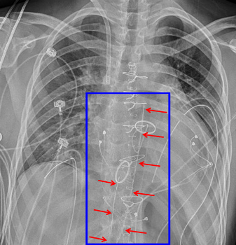
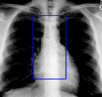
Median Sternotomy Wires
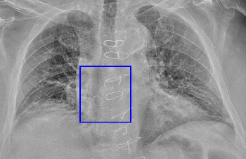
Wires used to close the surgical sternotomy, typically after cardiac or mediastinal surgery. Wires may tied in several fashions, e.g. cerclage, figure-of-eight. Very small wires are indicative of prior pediatric surgery, i.e. congenital heart disease.
Wires may fracture if the sternotomy is nonunited and there is motion. This is typically of no significance. However, in the immediate post-surgical setting wire fracture or wire migration can indicate sternal dehiscence.
|
|
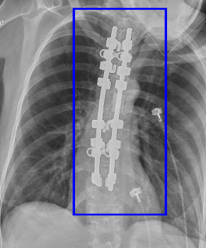
Spinal Fusion
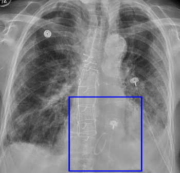
Generally involves rods (as seen here) as well as either pedicular screws or laminar hooks (the latter seen here) to provide initial fixation. Bone graft material, often difficult to see, provides long-term fusion of the involved levels.
|
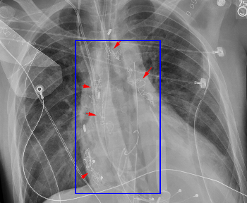
Surgical Gauze
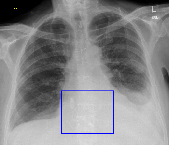
Surgical gauze is used for packing and contains radiopaque markers to help identify it on radiographs. It may be left in an open wound or retained inadvertently.
This patient with an open sternotomy has multiple gauzes and a single sponge in the lower chest.
|
