|
Central Venous Catheter
|
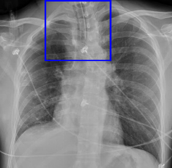
Denver Shunt
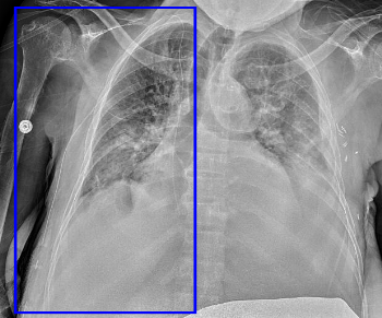
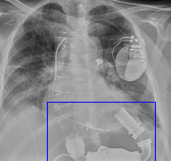
aka peritoneovenous shunt. Takes ascites to the venous system (generally internal jugular vein), traversing the chest wall. The radiolucent portion is the one-way valve.
|
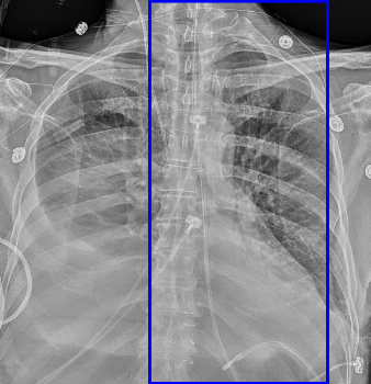
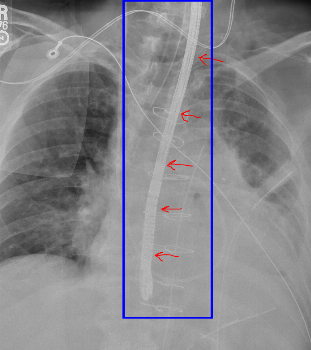
Esophageal Doppler Probe
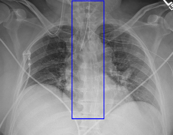
Used for non-invasive hemodynamic monitoring. Note the distinctive rectangular tip.
|
|
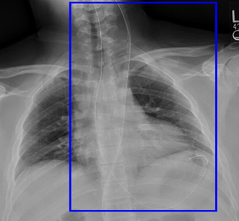
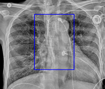
Esophageal Temperature Probe
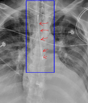
Braided-wire probe with radiopaque tip providing internal temperature monitoring.
|
Hemodialysis Catheter
Dual lumen central venous catheter, often tunneled, for short term dialysis access. The two lumens terminate at slightly different positions - the proximal opening is generally used for inflow ("arterial") and the distal opening is used for blood return ("venous"). The distal lumen should ideally lie within the right atrium.
|
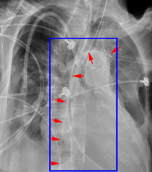
Left Ventricular Assist Device
Abbreviated LVAD. There are several different models of slightly varying size. The inflow cannula is generally radiopaque and lies at the left ventricular apex. The outflow cannula is generally radiolucent and plugs into the ascending aorta.
|
|
PICC
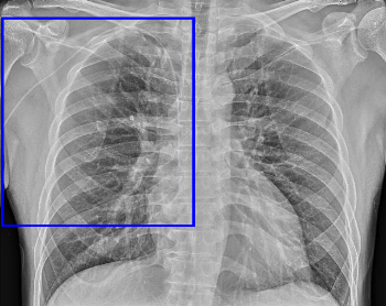
Peripherally inserted central venous catheter, used as an intermediate-term venous access. It is placed in the brachial or cephalic veins of the upper extremities (sometimes in the scalp or lower extremities in babies). The tip should ideally terminate at the superior cavoatrial junction.
Occasionally, the PICC will terminate within the axillary or subclavian vein, in which case it is referred to as a 'midline catheter' (since it is not central).
|
Port Catheter
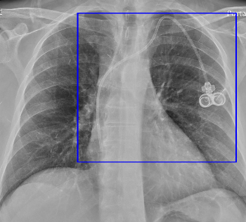
More permanent method of central venous access, consisting of a plastic/metal reservoir in the subcutaneous tissues and an intravenous catheter.
May be single lumen or, as in this case, dual lumen. Typically ports compatible with CT power injector will have a radiopaque "CT" marker on the reservoir.
|
Retained Guidewire
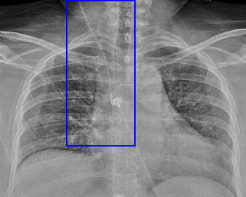
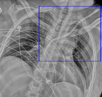
The guidewire used to place a central line using Seldinger technique may be inadvertently left behind after central line placement. These wires often have a 'J' tip, as in this case.
Note that this wire enters from the IVC, likely a femoral central line, and terminates in the left brachiocephalic vein.
|
|
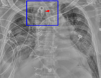
Transesophageal Echo Probe (TEE)
Used only intraoperatively to monitor cardiac surgery.
|
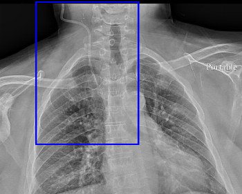
Ventriculoatrial Shunt
This may be indistinguishable from an internal jugular central venous catheter, although the high position in the neck might be a clue. This takes CSF from the ventricles into the SVC/right atrium.
|
