Biventricular Pacemaker
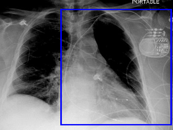
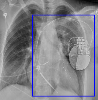
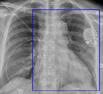
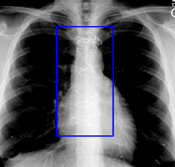
Leads in the right ventricle and a coronary vein (pacing the left ventricle). Typically these also have a right atrial lead, but they do not have to, such as in the example shown.
Denver Shunt
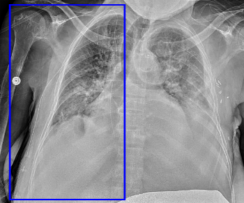
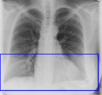
aka peritoneovenous shunt. Takes ascites to the venous system (generally internal jugular vein), traversing the chest wall. The radiolucent portion is the one-way valve.
Dual Chamber Pacemaker
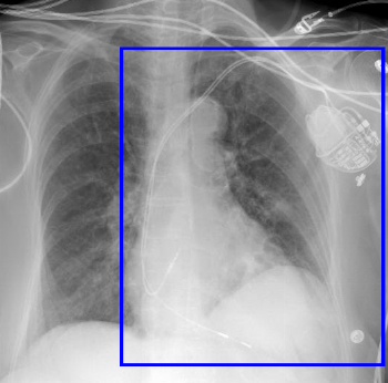
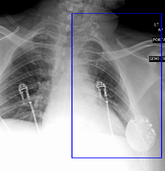
Two leads: right atrium and right ventricle.
Epicardial Pacemaker
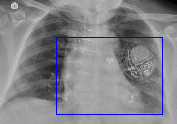
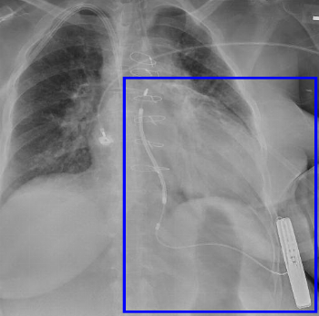
Epicardial leads are typically placed if transvenous access has failed. Temporary epicardial pacing leads are frequently used after cardiac surgery; however they are very much thinner and fainter on imaging.
Implanted Defibrillator
Implanted Cardioverter-Defibrillator (ICD) contains pacemaker and defibrillator functionality. It may have single chamber, dual chamber, or (often) biventricular leads. The leads in an ICD are characterized by one or more coils - a thickened portion that is used to deliver the shock. The pacemaker 'can' (pulse generator) is also distinguished by the presence of a capacitor as well as a battery.
The defibrillator component requires 2 coils; sometimes both coils are along a lead or leads in the venous system, but now the can itself can serve as one lead.
Inspire Sleep apnea device
The Inspire device is an upper airway neural stimulator device that has a sensor in the intercostal space for sensing breathing and a simulator in the neck for the hypoglossal nerve
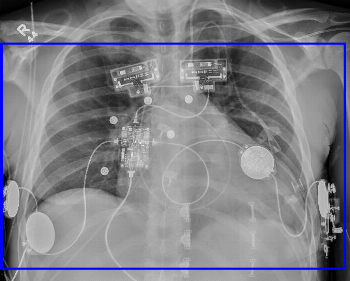
LifeVest
This is an entirely external vest worn by patients who cannot receive an implanted defibrillator.
Port Catheter
More permanent method of central venous access, consisting of a plastic/metal reservoir in the subcutaneous tissues and an intravenous catheter.
May be single lumen or, as in this case, dual lumen. Typically ports compatible with CT power injector will have a radiopaque "CT" marker on the reservoir.
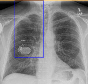
Single Chamber Pacemaker
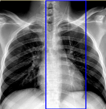
Single lead typically in the right ventricle.
Subcutaneous Defibrillator
A newer type of defibrillator, the subcutaneous defibrillator is entirely outside of the thoracic cage. The lead is placed anteriorly near the sternum, with the can along the lateral chest wall.
This can be particularly helpful in patients who need preserved central venous access, such as in this dialysis patient.
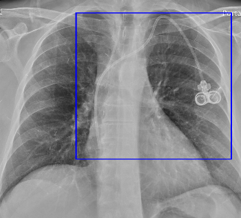
Vagal Nerve Stimulator
Used for patients with epilepsy. The lead goes to the supraclavicular fossa where the vagal nerve travels.
Ventriculoperitoneal Shunt
A VP shunt takes CSF from the ventricles to the peritoneal cavity. It generally courses along the anterior chest wall.
In this patient, there is one continuous catheter as well as an abandoned catheter fragment.
